Home
/ Animal Cell Parts Diagram : Structure Of An Animal Cell Diagram Illustration Of The Structure Of An Animal Cell Canstock / Animal cells depict various irregular shapes and sizes and are visible only under the microscope.
Animal Cell Parts Diagram : Structure Of An Animal Cell Diagram Illustration Of The Structure Of An Animal Cell Canstock / Animal cells depict various irregular shapes and sizes and are visible only under the microscope.
Animal Cell Parts Diagram : Structure Of An Animal Cell Diagram Illustration Of The Structure Of An Animal Cell Canstock / Animal cells depict various irregular shapes and sizes and are visible only under the microscope.. Labeled parts of a cell awesome plex eukaryotic plant cell. #animalcell #cell in this video i'm going to draw labelled diagram of animal cell.in this video you will see the diagram of animal cell and it's labelling. Unlike the eukaryotic cells of plants and fungi, animal cells do not have a cell wall. There are some differences between a plant and animal cell structure and functions. Animal cells are generally smaller than plant cells and lack a cell wall and chloroplasts;
Last updated on wed, 16 dec 2020 | medical terminology. Animal cell anatomy diagram structure with all parts nucleus. Diagram of animal cell anatomy stock illustration. Eukaryotic cell this is a. Describe the main parts of a cell.
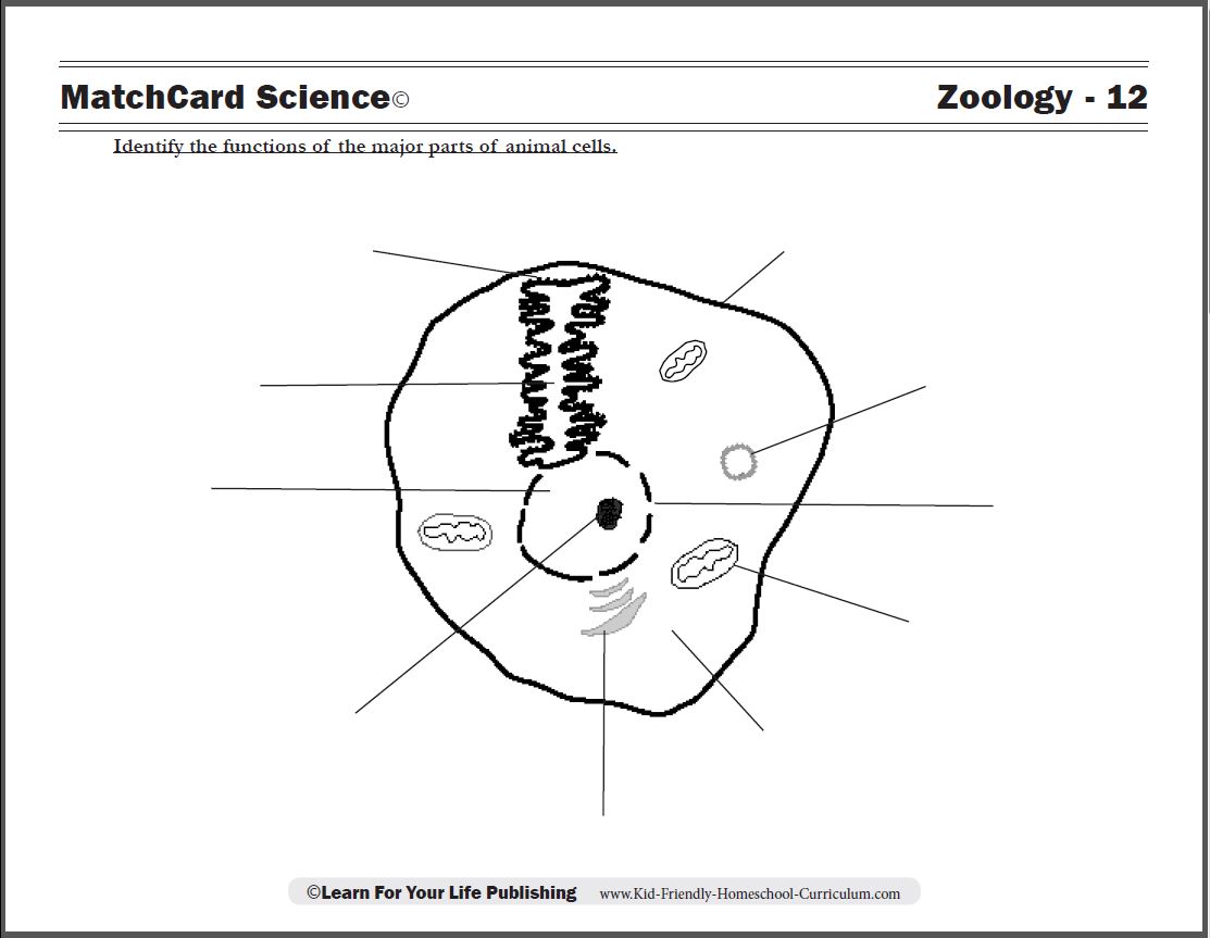
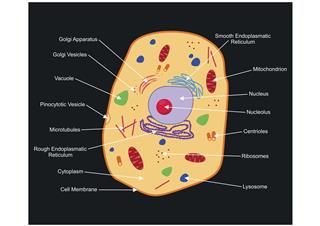
Animal Cell Diagram from www.learn4yourlife.com The centrosomes is where microtubules are made. Platelets help in blood clotting to prevent blood loss after injury. The animal cell and plant cell diagrams are easily colorable, allowing students to differentiate the different parts of the cell quickly. Cell organelles structure and parts. See how a generalized structure of an animal cell and plant cell look with labeled diagrams. Labeled parts of a cell awesome plex eukaryotic plant cell. The diagram, like the one above, will include labels of the major parts of an animal cell including the cell membrane, nucleus, ribosomes, mitochondria, vesicles, and cytosol. This pdf includes the color version, black and white version, and the labeled and unlabeled diagrams for students to complete.
Under the microscope, an animal cell shows many different parts called organelles, that work together to keep the cell functional.
Label a diagram of a typical cell. This diagram of animal cell is very important diagram for exam. After study of this chapter you should be able to: Parts of an animal cell biography. q in the animal cell diagram shown below, which part is pointing to the part that creates the spindle, the structure that separates chromosomes during cell division? Diagram showing anatomy of animal cell illustration royalty free. Here is a summary of their structure and function. Animal cell anatomy diagram structure with all parts nucleus. Label parts and thousands of other science skills. Cell is a tiny structure and functional unit of a living organism containing various parts known as organelles. The diagram clearly suggests er to be the second largest cell organelle after mitochondria since these form a series of interconnecting flattened tubular. Cells are the basic units of structure and function in living things. An animal cell diagram is a great way to learn and understand the many functions of an animal cell.
Animal cell structures, functions & diagrams. Animal cell diagrams labeled printable. Cell is a tiny structure and functional unit of a living organism containing various parts known as organelles. i this activity will improve your knowledge of cell parts and function. The diagram clearly suggests er to be the second largest cell organelle after mitochondria since these form a series of interconnecting flattened tubular.
A Labeled Diagram Of The Animal Cell And Its Organelles Biology Wise from pixfeeds.com Most notably, though using household electrical animal cell diagram labeled parts would not forget about one particular necessary saying. Diagram of an animal cell. A cell structure that forms a maze of passageways in which proteins and other materials are carried from one part of the cell to another. Diagram of animal cell anatomy stock illustration. Animal cell structure diagrammodel animal cell parts and. Animal cell parts and functions withcarbon. Animal cells and the membrane bound nucleus. These are organelles pertinent to plant cells.
Cell is a tiny structure and functional unit of a living organism containing various parts known as organelles.
Animal cell diagrams labeled printable. Animal cells and the membrane bound nucleus. Improve your science knowledge with free questions in animal cell diagrams: Label a diagram of a typical cell. Animal cells are generally smaller than plant cells and lack a cell wall and chloroplasts; The cell membrane, or plasma membrane, is a biological organelles are parts of the cell which are adapted and/or specialized for carrying out one or more vital functions. Cell organelles structure and parts. Keeping them on the same poster allows students to quickly understand the differences between the cells. This diagram of animal cell is very important diagram for exam. i this activity will improve your knowledge of cell parts and function. < structural biochemistry | cell organelles. Animal cell images stock photos vectors shutterstock. Let us look at animal cell parts and functions, using diagrams and illustrations.
Animal cell parts and functions withcarbon. The animal cell diagram on the worksheet identifies the major parts of the cell. Get the cell model powerpoints. After study of this chapter you should be able to: A vital part of an animal's immune system is white blood cell.
Plant And Animal Cells Ppt Video Online Download from slideplayer.com There are some differences between a plant and animal cell structure and functions. The cell is the basic functional and structural unit of life. Diagram showing anatomy of animal cell illustration royalty free. White blood cells help to kill damaging bacteria and other compounds which are harmful to the animal body. Animal cell structure diagrammodel animal cell parts and. Another important part of the cell is the cytoplasm. h cell parts and functions: Matchcard information pieces identify ten cellular parts and their function which are matched to the animal cell diagram.
At the center of the cell is the cell nucleus which contains the genetic code (dna).
Label a diagram of a typical cell. An animal cell is a type of eukaryotic cell that dominates most of the tissue cells in animals. After study of this chapter you should be able to: Animal cell anatomy diagram structure with all parts nucleus. This pdf includes the color version, black and white version, and the labeled and unlabeled diagrams for students to complete. The animal cell and plant cell diagrams are easily colorable, allowing students to differentiate the different parts of the cell quickly. Animal cells depict various irregular shapes and sizes and are visible only under the microscope. These are organelles pertinent to plant cells. Keeping them on the same poster allows students to quickly understand the differences between the cells. Under the microscope, an animal cell shows many different parts called organelles, that work together to keep the cell functional. Animal cells are all smooched. Animal cells are generally smaller than plant cells and lack a cell wall and chloroplasts; This diagram of animal cell is very important diagram for exam.
Share :
Post a Comment
for "Animal Cell Parts Diagram : Structure Of An Animal Cell Diagram Illustration Of The Structure Of An Animal Cell Canstock / Animal cells depict various irregular shapes and sizes and are visible only under the microscope."


Post a Comment for "Animal Cell Parts Diagram : Structure Of An Animal Cell Diagram Illustration Of The Structure Of An Animal Cell Canstock / Animal cells depict various irregular shapes and sizes and are visible only under the microscope."